Optical Coherence Tomography (OCT): High-resolution imaging for detailed analysis of the retina, macula, and optic nerve head
Fundus Photography & Fluorescein Angiography (FFA): Advanced retinal imaging for vascular disorders, diabetic retinopathy, and macular diseases
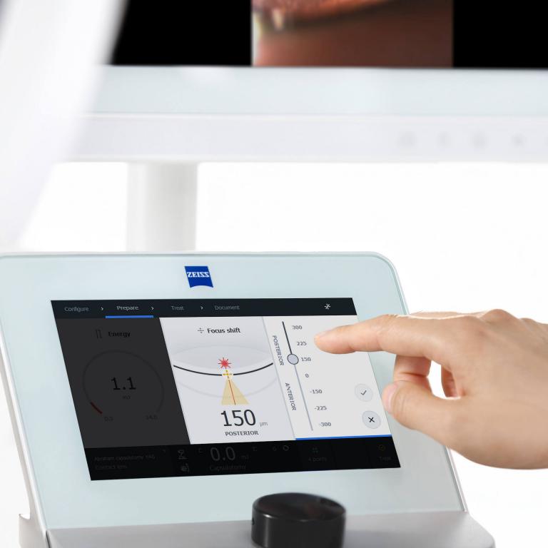
Corneal Topography & Tomography (Pentacam / Atlas): 3D mapping of the cornea for keratoconus detection, refractive surgery planning, and corneal diseases
Corneal Pachymetry: Accurate corneal thickness measurement for glaucoma risk assessment and LASIK suitability
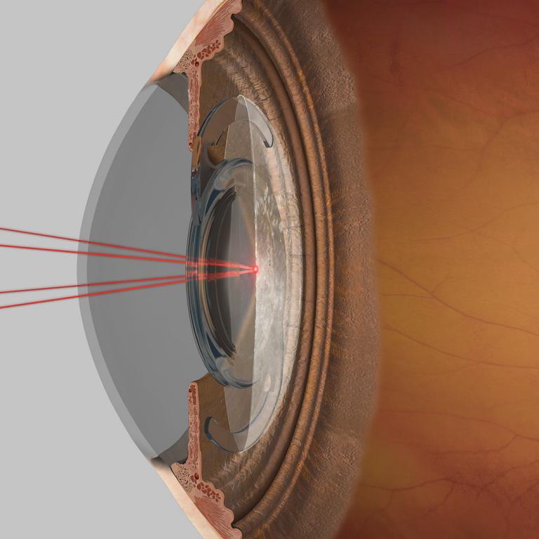
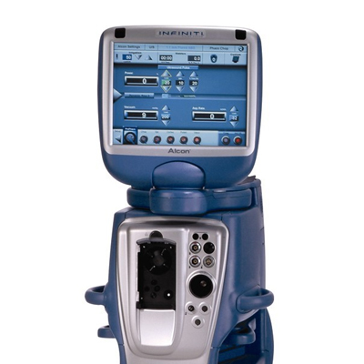
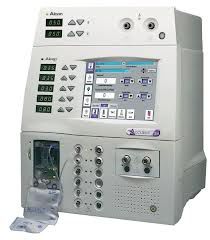
Optical Biometry (IOL Master, Alcon Ocuscan): Precision calculation of intraocular lens power for cataract and refractive surgeries
A-Scan & B-Scan Ultrasonography: For ocular dimensions and posterior segment evaluation in dense cataracts or vitreous hemorrhage
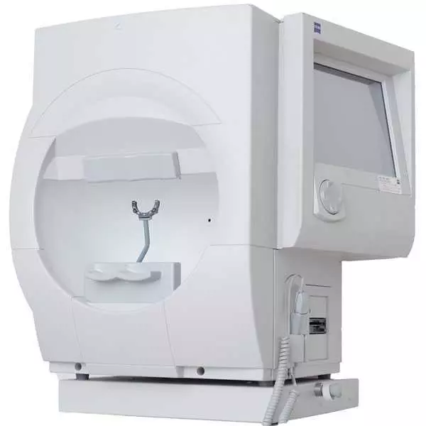
Visual Field Analysis (Humphrey Visual Field): For detection, staging, and monitoring of glaucoma and optic neuropathies
tonometry (Applanation & Non-Contact): Reliable intraocular pressure measurement to diagnose and manage glaucoma
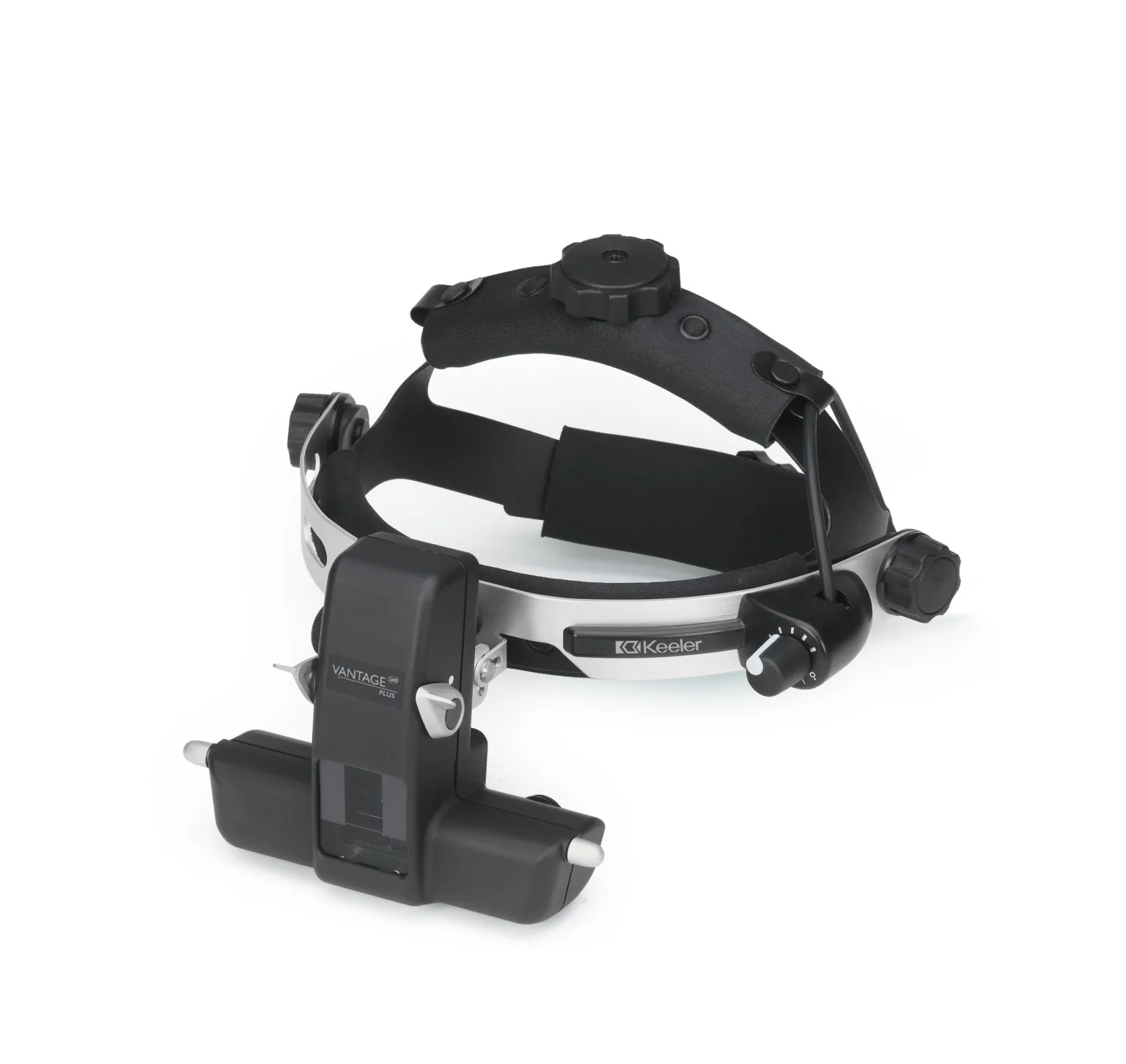
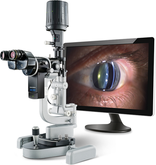
Anterior Segment Imaging (AS-OCT, Specular Microscopy): Detailed analysis of corneal endothelium, anterior chamber depth, and angle structures
Electrodiagnostic Testing (ERG, VEP, EOG): For functional assessment of the retina, optic nerve, and visual pathways
Orthoptic & Binocular Vision Assessment: For squint (strabismus), amblyopia, and pediatric ophthalmology evaluation
Dry Eye Evaluation (Tear Film Breakup Time, Schirmer’s Test): For diagnosis and monitoring of ocular surface disorders
Color Vision & Contrast Sensitivity Testing: Essential for optic nerve diseases and macular pathologies
Endothelial Cell Count Analysis: For corneal health assessment, especially before corneal transplants and in dystrophies
Ocular Biometry & Axial Length Measurement: Critical for refractive and cataract surgical planning
Wavefront Aberrometry: For detection of higher-order aberrations in refractive surgery candidates
Ultrasound Biomicroscopy (UBM): High-frequency ultrasound for detailed imaging of anterior segment structures
Confocal Microscopy: For in-vivo analysis of corneal microstructures in infections and dystrophies